Goat Eyelid: A Deep Dive into Anatomy, Function, and Welfare
Goats, with their charming personalities, insatiable curiosity, and often mischievous glint in their eyes, are a beloved species of livestock worldwide. Yet, beneath their seemingly simple appearance lies a complex and fascinating array of biological adaptations, many of which contribute to their remarkable resilience and ability to thrive in diverse environments. Among these, the goat eyelid, often overlooked, plays a crucial role in the animal’s overall health, sensory perception, and even its unique communication. This extensive article will delve into the intricate world of the goat eyelid, exploring its detailed anatomy, multifaceted functions, common ailments, and the vital importance of proper care and welfare.
A Masterpiece of Evolution: Anatomy of the Goat Eyelid
To truly appreciate the significance of the goat eyelid, we must first understand its structural components. Unlike humans who possess a relatively simple eyelid structure, goats, like many ungulates, exhibit a more complex design, finely tuned for their specific needs.
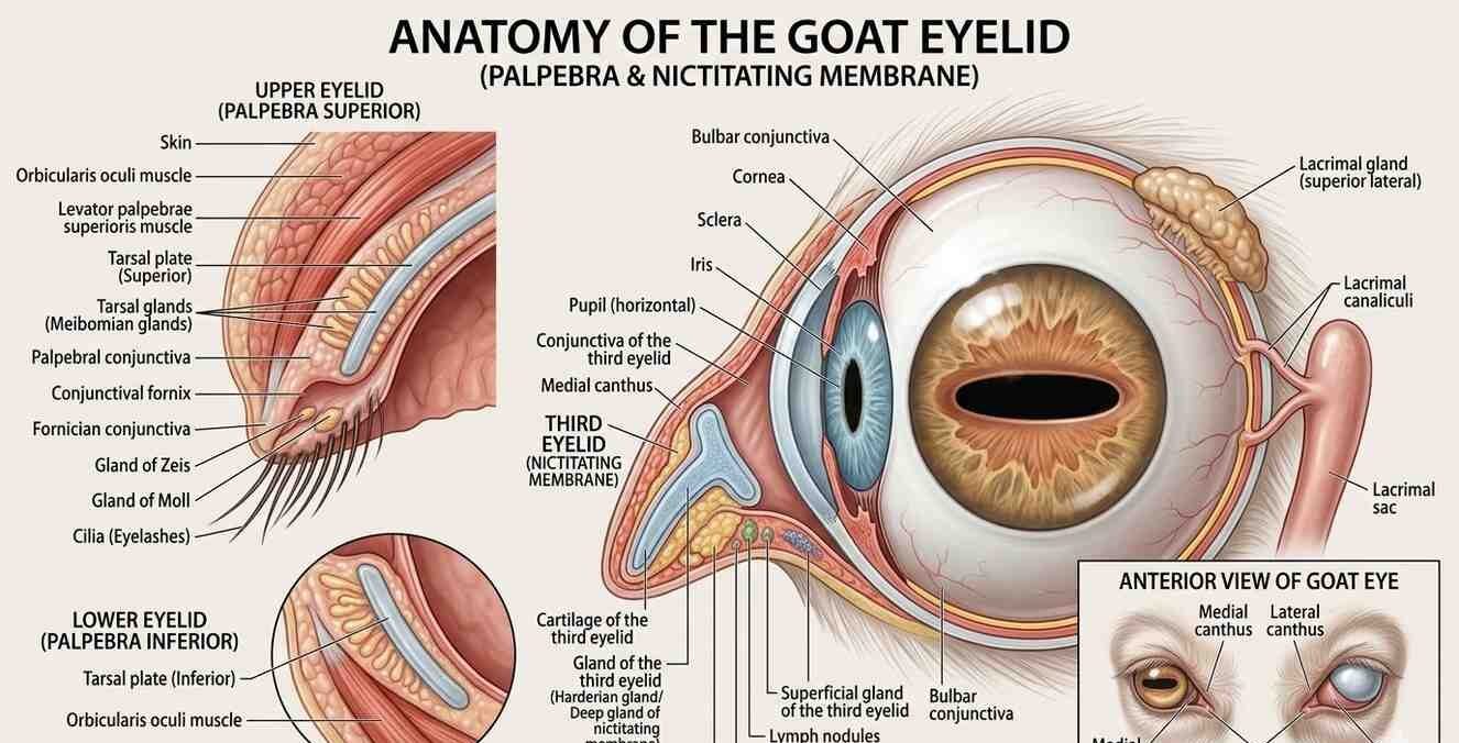
1. The Upper and Lower Lids: At the most fundamental level, the goat possesses distinct upper and lower eyelids. These are fleshy folds of skin, supported by a combination of connective tissue, muscle, and a specialized cartilaginous plate known as the tarsal plate. The tarsal plate provides rigidity and shape to the eyelids, preventing them from collapsing and ensuring a smooth, effective blink. The skin covering the eyelids is thin and pliable, allowing for a wide range of movement.
2. The Orbicularis Oculi Muscle: The primary muscle responsible for closing the eyelids is the orbicularis oculi. This circular muscle surrounds the eye and, upon contraction, draws the eyelids together, facilitating blinking and tightly sealing the eye for protection. Its efficient action is paramount for quick responses to environmental threats.
3. The Levator Palpebrae Superioris Muscle: While not directly part of the eyelid itself, the levator palpebrae superioris muscle plays a critical role in eyelid function by elevating the upper eyelid. This allows for the widening of the palpebral fissure (the opening between the eyelids), granting the goat a broad field of vision.
4. Eyelashes (Cilia): Perhaps the most visually striking feature of the goat eyelid are its eyelashes. These short, stiff hairs project from the margins of both the upper and lower eyelids. Far from being merely decorative, eyelashes serve as a vital protective barrier, filtering out dust, debris, and insects before they can reach the delicate surface of the eyeball. They act as a sophisticated early warning system, triggering a blink reflex when foreign objects make contact.
5. Meibomian Glands (Tarsal Glands): Embedded within the tarsal plates of both eyelids are modified sebaceous glands called Meibomian glands. These glands produce a lipid-rich secretion known as meibum. Meibum forms the outermost layer of the tear film, preventing rapid evaporation of the watery tear layer and ensuring a stable, lubricated ocular surface. A healthy meibomian gland function is crucial for preventing dry eye syndrome.
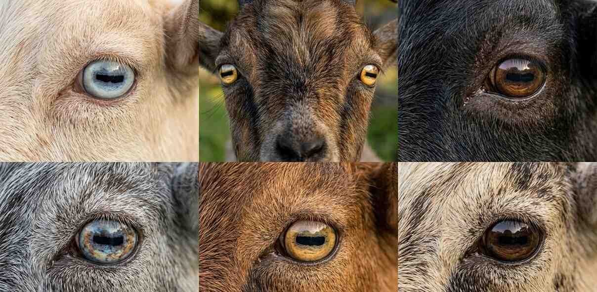
6. Conjunctiva: Lining the inner surface of the eyelids and reflecting onto the anterior surface of the eyeball (up to the limbus, where it meets the cornea) is the conjunctiva. This delicate mucous membrane is highly vascularized and plays a vital role in immune defense, producing components of the tear film, and facilitating smooth movement of the eyeball within the orbit. The bulbar conjunctiva, covering the eyeball, is typically clear, allowing the underlying sclera (white of the eye) to be visible. The palpebral conjunctiva, lining the eyelids, is often pinkish.
7. The Nictitating Membrane (Third Eyelid): One of the most distinctive features of the goat’s ocular anatomy, shared with many other animals but absent in humans, is the nictitating membrane, often referred to as the “third eyelid.” Located in the medial (inner) corner of the eye, this translucent, T-shaped fold of conjunctiva and cartilage can rapidly sweep across the surface of the cornea. Its primary functions are multifaceted:
Protection: It provides an extra layer of defense against dust, foreign bodies, and strong winds, particularly important for grazing animals that frequently lower their heads.
Tear Film Distribution: It helps to spread the tear film evenly across the cornea, ensuring optimal lubrication and hydration.
Gland of the Third Eyelid: Associated with the nictitating membrane is the gland of the third eyelid (Harderian gland), which contributes significantly to tear production, particularly the serous (watery) component.
Understanding these anatomical components provides a foundation for appreciating the complex interplay of structures that contribute to the remarkable efficiency of the goat eyelid.
More Than Just a Blink: Functions of the Goat Eyelid
The goat eyelid is a marvel of biological engineering, performing a wide array of critical functions essential for the animal’s vision, health, and survival.
1. Ocular Protection: This is arguably the most paramount function of the eyelids. Goats are prey animals and forage in varied, often challenging environments. Their eyes are constantly exposed to dust, dirt, thorns, branches, insects, and harsh weather conditions. The eyelids act as a robust physical barrier, rapidly closing to shield the delicate cornea and conjunctiva from potential harm. The blink reflex, triggered by sensory input, ensures swift protection.
2. Tear Film Maintenance and Distribution: The tear film is a complex three-layered fluid that constantly bathes the surface of the eye. It is crucial for:
Lubrication: Reducing friction between the eyelids and the eyeball during blinking.
Hydration: Keeping the corneal and conjunctival cells moist and healthy.
Nutrient Supply: Delivering oxygen and nutrients to the avascular (lacking blood vessels) cornea.
Waste Removal: Flushing away debris and metabolic byproducts.
Antimicrobial Defense: Containing antibodies and enzymes that fight off infections. The eyelids, through their blinking action and the contributions of the Meibomian glands and the gland of the third eyelid, are integral to producing, distributing, and maintaining the integrity of this vital tear film.
3. Vision Optimization: While primarily protective, the eyelids also play an indirect role in optimizing vision. By clearing the corneal surface of debris and maintaining a smooth tear film, they ensure that light can enter the eye unimpeded, allowing for clear image formation on the retina. A compromised tear film or irritated eyelids can lead to blurred vision, impacting the goat’s ability to forage and detect predators.
4. Light Regulation: In bright sunlight, goats can partially narrow their eyelids, acting like a natural aperture to reduce the amount of light entering the eye. This helps prevent glare and photophobia (light sensitivity), allowing them to maintain comfortable vision even in intense conditions.
5. Communication and Expression: Though perhaps less overtly expressive than in humans, the goat’s eyelids, in conjunction with other facial muscles, contribute to subtle forms of communication and expression. A relaxed, open eye often indicates a calm and healthy animal, while squinting, excessive blinking, or drooping eyelids can signal pain, discomfort, or illness. Experienced handlers often observe these subtle cues to gauge the welfare of their animals.
When Eyelids Go Wrong: Common Ailments and Conditions
Despite their robust design, goat eyelids are susceptible to a range of ailments that can compromise their function and impact the animal’s welfare. Early detection and appropriate treatment are crucial for preventing more serious complications.
1. Eyelid Trauma and Lacerations: Given their active nature and propensity for exploring thorny bushes or engaging in playful headbutting, goats are prone to eyelid trauma. Lacerations can range from superficial scratches to deep tears, potentially affecting the integrity of the eyelid margin, eyelashes, and even the lacrimal (tear) drainage system. Prompt veterinary attention is necessary to clean the wound, assess the extent of the damage, and perform surgical repair if needed. Untreated lacerations can lead to scar tissue formation, entropion, or ectropion.
2. Entropion: Entropion is a genetic condition where a portion of the eyelid margin, typically the lower lid, rolls inward towards the eye. This causes the eyelashes and surrounding hair to rub against the delicate cornea and conjunctiva, leading to irritation, pain, excessive tearing (epiphora), corneal ulcers, and potentially blindness if left untreated. Entropion is more commonly seen in young goats and certain breeds. Treatment often involves surgical correction to evert the eyelid margin.
3. Ectropion: Ectropion is the opposite of entropion, where the eyelid margin rolls outward, exposing the conjunctiva. This can lead to chronic dryness, inflammation, and increased susceptibility to foreign bodies and infections, as the protective function of the eyelid is compromised. While less common in goats than entropion, it can occur due to trauma, scarring, or certain neurological conditions. Treatment focuses on addressing the underlying cause and may involve surgical correction.
4. Conjunctivitis (Pinkeye): Conjunctivitis, or inflammation of the conjunctiva, is a common ocular ailment in goats. It can be caused by various factors, including bacterial infections (e.g., Chlamydophila, Mycoplasma, Moraxella), viral infections, allergens, or irritants (dust, ammonia fumes). Symptoms include redness, swelling of the conjunctiva, ocular discharge (ranging from watery to thick and purulent), squinting, and photophobia. Infectious conjunctivitis can be highly contagious within a herd. Treatment depends on the underlying cause and may involve topical antibiotics, anti-inflammatory drugs, or antiviral medications.
5. Keratoconjunctivitis (Often “Pinkeye”): When conjunctivitis progresses to involve the cornea (keratitis), it becomes keratoconjunctivitis. This is often seen in severe cases of infectious pinkeye, particularly those caused by Mycoplasma species. Corneal involvement can lead to cloudiness, ulcers, and potentially permanent vision impairment if not treated aggressively.
6. Blepharitis: Blepharitis refers to inflammation of the eyelids themselves, particularly the eyelid margins. It can be caused by bacterial infections, parasitic infestations (mites), allergies, or irritation. Symptoms include redness, swelling, scaling, crusting along the eyelid margins, and itching. Treatment involves identifying and addressing the underlying cause, often with topical medications and meticulous hygiene.
7. Ocular Foreign Bodies: Given their grazing habits, goats are prone to getting foreign bodies lodged in their eyes. Small pieces of hay, seeds, thorns, or dust can become trapped under the eyelids or in the conjunctival sac, causing severe irritation, pain, tearing, and potential corneal damage. If a foreign body is suspected, it is crucial to carefully examine the eye, including everting the eyelids and checking under the third eyelid. Veterinary assistance may be required for removal, especially if the object is embedded or causing significant pain.
8. Abscesses and Cysts: Bacterial infections can sometimes lead to the formation of abscesses within or around the eyelid tissues. These painful, pus-filled swellings require drainage and antibiotic treatment. Cysts, often arising from blocked Meibomian glands (chalazion) or other glandular structures, can also occur and may require surgical removal if they become problematic.
9. Tumors: While less common, goats can develop tumors of the eyelids or surrounding ocular structures. These can be benign or malignant and may manifest as lumps, bumps, or changes in eyelid shape or texture. Any suspicious growth should be examined by a veterinarian for diagnosis and appropriate treatment, which may include surgical excision.
The Cornerstone of Health: Eyelid Care and Welfare
Maintaining the health of a goat’s eyelids is an integral part of responsible animal husbandry. Proactive measures and vigilant observation can prevent many problems and ensure the animal’s comfort and optimal vision.
1. Regular Observation: Routine daily observation of your goats is paramount. Pay close attention to their eyes:
2. Environmental Management: The goat’s living environment significantly impacts eyelid health.
Dust Control: Minimize dust in barns and feeding areas. Use dust-extracted bedding and ensure good ventilation.
Ammonia Levels: High ammonia levels from urine and feces can irritate the eyes. Ensure adequate ventilation and regular cleaning of housing.
Protection from Hazards: Remove sharp objects, protruding wires, or thorny plants from grazing areas and enclosures that could cause eyelid trauma. Provide shelter from strong winds and harsh sunlight.
Fly Control: Flies can transmit infectious agents and cause irritation around the eyes. Implement fly control measures, such as traps, repellents (safe for goats), and good sanitation.
3. Nutritional Support: A well-balanced diet rich in essential vitamins and minerals is crucial for overall health, including ocular health. Deficiencies in certain vitamins (e.g., Vitamin A) can sometimes manifest as ocular problems.
4. Hygiene: Maintain good hygiene around the eyes, especially if discharge is present. Gently clean away any crusts or discharge with a clean cloth dampened with warm water or a veterinarian-recommended ophthalmic solution. Always use separate cloths for each eye and each animal to prevent the spread of infection.
5. Prompt Veterinary Intervention: If you notice any signs of eyelid or ocular problems, contact your veterinarian immediately. Early diagnosis and treatment are critical for preventing complications and preserving vision. Self-treating can often exacerbate the problem or delay appropriate medical care.
6. Biosecurity: If you introduce new goats to your herd, quarantine them and observe them closely for any signs of illness, including ocular issues, before integrating them with the main herd. This helps prevent the spread of infectious eye diseases.
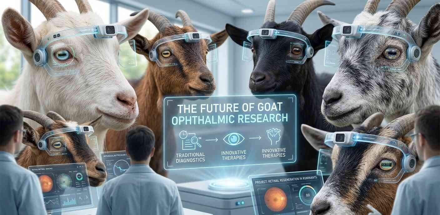
The Future of Goat Ophthalmic Research
As our understanding of goat physiology and health continues to expand, so too does research into specific areas like ophthalmic health. Future research may focus on:
Genetic predispositions: Identifying specific genetic markers for conditions like entropion to aid in selective breeding programs.
Advanced diagnostics: Developing more sophisticated diagnostic tools for early detection of ocular diseases. Novel treatments: Exploring new pharmacological agents or surgical techniques for managing complex eyelid and eye conditions.
Environmental impacts: Further studying the long-term effects of various environmental stressors on goat eye health.
Conclusion: A Small Structure, A Big Impact
The goat eyelid, though seemingly a minor anatomical feature, is in reality a sophisticated and indispensable component of the animal’s biology. Its intricate structure and multifaceted functions are crucial for ocular protection, tear film maintenance, and overall visual health. From the protective sweep of the nictitating membrane to the subtle warnings of the eyelashes, every part of the eyelid plays a vital role in enabling goats to navigate their world safely and effectively.